Friday, January 29, 2016

Thyroid Disfunction by Flouride toxicity

THYROID

The thyroid gland regulates the body’s metabolic rate and plays an exquisitely important role in human health. Because all metabolically active cells require thyroid hormone for proper functioning, thyroid disruption can have a wide range of effects on virtually every system of the body. Chemicals that interfere with thyroid function must be treated with great caution. According to the U.S. National Research Council, and as discussed below, there is substantial evidence that fluoride exposure can impact thyroid function in some individuals. (NRC 2006).

Fluoride Was Once Prescribed as an Anti-Thyroid Drug

When people think of fluoride being prescribed for medicinal purposes, they generally think of fluoride supplementation to reduce tooth decay. Fluoride, however, has also been prescribed as a drug to reduce the activity of the thyroid gland. Up through the 1950s, doctors in Europe and South America prescribed fluoride to reduce thyroid function in patients with over-active thyroids (hyperthyroidism). (Merck Index 1968). Doctors selected fluoride as a thyroid suppressant based on findings linking fluoride to goitre, and, as predicted, fluoride therapy did reduce thyroid activity in the treated patients. (McClaren 1969; Galletti 1958; May 1937). Moreover, according to clinical research, the fluoride dose capable of reducing thyroid function was notably low — just 2 to 5 mg per day over several months. (Galletti & Joyet 1958). This dose is well within the range (1.6 to 6.6 mg/day) of what individuals living in fluoridated communities are now estimated to receive on a regular basis. (DHHS 1991).

Fluoride & Hypothyroidism

Based on fluoride’s anti-thyroid effects in hyperthyroid patients, concerns have arisen about whether current fluoride exposures could be contributing to the increased prevalence of under-active thyroid (clinical and/or subclinical hypothyroidism) in the United States and other nations. In February 2015, British scientists reported that fluoridated water in Britain is associated with elevated rates of hypothyroidism:

“We found that higher levels of fluoride in drinking water provide a useful contribution for predicting prevalence of hypothyroidism. We found that practices located in the West Midlands (a wholly fluoridated area) are nearly twice as likely to report high hypothyroidism prevalence in comparison to Greater Manchester (non-fluoridated area).” (Peckham 2015).

Supporting the fluoride/hypothyroidism connection are a number of studies from China, India, and Russia that have found alterations in thyroid hormones, including reduced T3 and increased TSH, in populations exposed to elevated levels of fluoride in the workplace or in the water. (NRC 2006; Susheela 2005; Mikhailets 1996; Yao 1996; Bachinskii 1985; Yu 1985).

In clinical hypothyroidism, the thyroid gland fails to produce sufficient quantities of the hormones triiodothyronine (T3) and thyroxine (T4). These hormones are required by all metabolically active cells, and their reduced presence can thus produce a range of ill effects, including fatigue, muscle/joint pain, depression, weight gain, menstrual disturbances, impaired fertility, impaired memory, and inability to concentrate. When T3 and T4 levels begin to fall, the pituitary gland responds by increasing production of “Thyroid Stimulating Hormone” (TSH) as a means of getting the thyroid to produce more T3 and T4.

In subclinical hypothyroidism, the TSH level is elevated, but the T3 and T4 hormones are still within the normal range. Although subclinical hypothyroidism used to be regarded as largely inconsequential, it is increasingly considered a “clinically important disorder.” (Gencer 2012). Some studies have found, for example, that subclinical hypothyroidism in pregnant women results in reduced IQ in offspring, (Klein 2001; Haddow 1999), and a recent study in the Journal of the American Medical Association found that adults with subclinical hypothyroidism had a significantly higher rate of coronary heart disease. (Rodondi 2010).

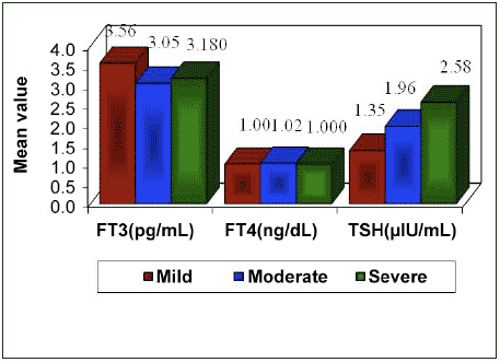
Thyroid Hormone Levels Based on Severity of Dental Fluorosis (Hosur 2012).

Studies investigating fluoride’s impact on thyroid hormone levels have produced divergent findings, but are consistent with fluoride having an anti-thyroid effect under certain circumstances. (NRC 2006). The most common thyroid effect associated with fluoride exposure appears to be an increase in TSH levels, with or without a corresponding effect on T3 or T4. (Susheela 2005). One of the most recent studies, for example, found a trend towards higher TSH in children based on the severity of their dental fluorosis, but without a significant effect on either T3 or T4. (Hosur 2012, see figure). These and other findings indicate that fluoride can contribute to a subclinical, if not clinical, hypothyroid condition. It remains difficult to predict the toxic dose, however, as it appears to depend, in part, on the nutritional and health status of the individual, particularly the adequacy of iodine intake. (NRC 2006).

Fluoride Exacerbates the Impact of Iodine Deficiency

A consistent body of animal and human research shows that fluoride exposure worsens the impact of an iodine deficiency. (Gas’kov 2005; Hong 2001; Wang 2001; Zhao 1998; Xu 1994; Lin 1991; Ren 1989; Guan 1988). Iodine is the basic building block of the T3 and T4 hormones and thus an adequate iodine intake is essential for the proper functioning of the thyroid gland. When iodine intake is inadequate during infancy and early childhood, the child’s brain can suffer permanent damage, including mental retardation. (Iodine deficiency is the leading cause of mental retardation throughout the world.)

In China, researchers have repeatedly found that an iodine deficiency coupled with fluoride exposure produces a significantly more damaging effect on neurological development than iodine deficiency alone. (Hong 2001; Xu 1994; Lin 1991; Ren 1989). The studies, which utilize childhood intelligence as the metric for assessing neurological health, have found that fluoride levels as low as 0.9 ppm can worsen the IQ effect of iodine deficiency. (Lin 1991). This concentration is within the purported “optimal” range of fluoride that is added to water in water fluoridation programs (0.7-1.2 ppm). While many studies have found an association between fluoride and reduced IQ among children with adequate iodine intake, (Choi 2012), an iodine deficiency will lower the threshold at which fluoride damages the brain. (Xu 1994; Guan 1988). An iodine deficiency will also lower the threshold for other forms of fluoride toxicity as well, including dental fluorosis. (Zhao 1998; see also Pontigo-Loyola 2008).

Iodine Deficiency Remains a Public Health Concern in the U.S.

Despite the widespread availability of iodized salt, iodine deficiency has re-emerged as a public health concern in the United States. (CDC 1998). More than 11% of all Americans, and more than 15% of American women of child-bearing age, presently have urine iodine levels less than 50 mcg/L (Caldwell et al., 2008), indicating moderate to severe iodine deficiency. An additional 36% of reproductive-aged women in the U.S. are considered mildly iodine deficient (<100 mcg/L urinary iodine). Fluoride’s ability to worsen the effects of an iodine deficiency could thus be highly relevant to populations in the U.S. The National Research Council has therefore called upon the scientific community to begin investigating the interactive effects of fluoride and iodine in U.S. populations. So far, no such research has been conducted.

Fluoride & Goitre

Studies dating back to the 19th century have implicated fluoride as a possible cause of goitre. Goitre (aka goiter) is an enlargement of the thyroid gland that in some cases can produce visible swelling in the neck. Although the main cause of goitre is iodine deficiency, it can also be caused by other things, including hypothyroidism and goitrogens (substances that cause goitre). Studies that have examined human populations with adequate intake of iodine have reported mixed results about fluoride’s ability to produce goitre. (NRC 2006; Burgi 1984; McLaren 1969). The research has been more consistent, however, where the examined populations had either excessive iodine intakes, or deficient iodine intakes. (Gas’kov 2005; Hong 2001; Wang 2001; Xu 1994; Yang 1994; Lin 1986). Most of this latter research was initially published in either Russian or Chinese and was only recently translated into English by the Fluoride Action Network. Accordingly, previous reviews of fluoride/goitre research (e.g., NRC 2006) were not able to take these studies into account. As such, the evidence linking fluoride to goitre for populations with excessive, or deficient, iodine exposure is stronger than previously recognized. Read more.

Fluoride, Thyroid, & Dogs

An investigation by the Environmental Working Group found that commercial dog food contains very high levels of fluoride (due, in part, to the presence 0f fluoride-rich bone particles). Since dogs have been found to suffer a high incidence of hypothyroidism, the relationship between fluoride contamination and thyroid disease in dogs deserves further attention, particularly since it was fluoride’s production of goiter in dogs that first prompted the idea that fluoride could be an anti-thyroid agent. (Maumene 1854).

REFERENCES:

back to top

    Wednesday, December 16, 2015

    How to Think About the Risk of Autism

    Sunday Review New York Times.  

    OPINION

    How to Think About the Risk of Autism

    By SAM WANG

    STUDY published last week found that the brains of autistic children show abnormalities that are likely to have arisen before birth, which is consistent with a large body of previous evidence. Yet most media coverage focuses on vaccines, which do not cause autism and are given after birth. How can we help people separate real risks from false rumors?

    Over the last few years, we’ve seen an explosion of studies linking autism to a wide variety of genetic and environmental factors. Putting these studies in perspective is an enormous challenge. In a database search of more than 34,000 scientific publications mentioning autism since its first description in 1943, over half have come since 2008.

    As a statistically minded neuroscientist, I suggest a different approach that relies on a concept we are familiar with: relative odds. As a single common measuring stick to compare odds, I have chosen the “risk ratio,” a measure that allows the bigger picture to come into focus.

    For a variety of studies I asked the same question: How large is the increased risk for autism? My standard for comparison was the likelihood in the general population of autism spectrum disorder. Here’s an example. Start from the fact that the recorded rate of autism is now 1 in 68, according to a report released last week by the Centers for Disease Control and Prevention. If babies born in purple farmhouses have a rate of autism of 2 in 68, this doubling means that the purple farmhouse carries a risk ratio of 2. However, correlation is not causation, and there is no need to repaint that farmhouse just yet.

    We can improve our chances of finding true causes by looking before the age of 2, when it becomes possible to diagnose autism. The risk ratio can give perspective where isolated news stories don’t. Media reports have focused on the risk associated with becoming a mother or father in one’s late 30s or after. The story has obvious appeal: Delayed parenthood is common, and readers are understandably anxious. However, parents-to-be should consider that the individual risk to the child is only around 1.4. The risk associated with enhanced or accelerated labor in full-term babies is about 1.2, after other complications are taken into account. And of course, the risk from vaccination is slightly less than 1 — there is no added risk. Even worse, incorrect beliefs about vaccines come with a cost. The return of measles in communities with falling vaccination rates is one recent example.

    One might argue that any added risk is too much. But it is essential to put risk into perspective. The early research in the 1950s that first linked smoking with lung cancer gave a risk ratio around 25 for a pack-a-day smoker. In the case of autism, the risk associated with parental age is dwarfed by the impact of inheritance.

    Comparing twins who grow up in the same environment allows researchers to focus on the effects of shared genes. A majority of children with an autistic identical twin are likely to be diagnosed with autism as well for a risk ratio somewhere around 80. A lower risk ratio comes from sharing half of one’s genes, as for fraternal twins or siblings. For these reasons, researchers believe that autism’s roots are largely genetic.

    The human genome is dotted with hundreds of autism risk genes. Individually, each gene is usually harmless, and is in fact essential for normal function. This suggests that autism is caused by combinations of genes acting together. Think of genes as being like playing cards, and think of autism outcomes as depending on the entire hand. New mutations can also arise throughout life, which might account for the slightly greater risk associated with older fatherhood.

    Although autism has a gene-based beginning, growing brains are also influenced by their environment and external events. Looking at when these risks are greatest can provide clues about when the growing brain is most vulnerable. Based on a large body of evidence, the known hazards occur before birth and fall into three broad categories: prematurity, prenatal stress and brain development.

    MANY known risks for autism occur during late pregnancy and birth. Premature birth is a risk for developmental disability, including autism. Notably, elective cesarean section is associated with an autism risk ratio of 1.9. Since a substantial proportion of early deliveries are elective, without a compelling medical reason, this risk is preventable.

    A highly underappreciated prenatal risk is stress. For pregnant women who take the sometimes-wrenching step of emigrating to a new country, for example, the risk ratio is 2.3. In the fifth through ninth months of pregnancy, getting caught in a hurricane strike zonecarries a risk ratio of about 3. Maternal post-traumatic stress disorder during pregnancy is associated with a similar effect. These events are likely to trigger the secretion of stress hormones, which can enter the fetus’s bloodstream and affect the developing brain for a lifetime. Stressors may also lead to maternal illness, the immune response to which may interfere with brain development.

    Stress might account for other findings as well. Recent news coverage has speculated on the influence of air pollutants, which carry risk ratios around 1.4. This risk might be caused by chemicals — or by the stress of living in a poor or crowded neighborhood, where pollution is worse. A larger risk comes from households that already have an older sibling under 1 year of age, where newly conceived children have a risk ratio for autism of 3.4. So sure, parents should avoid smog — but also might think about spacing their children at judiciously chosen intervals.

    After birth, known risks diminish. But the baby’s brain acquires a new need: social experience. In one group of Romanian orphanage children, babies were nearly isolated from social contact, and some later showed autism-like symptoms. Developing brains go through sensitive periods during which they require a minimum level of normal experience. Extreme deprivation may affect a critical period of the brain’s social and emotional development.

    Risk ratios are good not just for parents, but also for researchers, who can follow them to new areas for in-depth study. The statistics suggest a conceptual framework in which fetal brain development stays on track unless it is driven awry by genetic hits, by adversity in late pregnancy, or a combination of the two.

    In my laboratory, risk ratios have led us to examine particular brain regions. Many brain regions in autistic people show abnormalities, but it is not known whether some malfunctioning regions cause other regions to go off track. In one study focusing on detailed long-term outcomes in 51 children, damage to the cerebellum at birth leads to a risk ratio of about 40. The cerebellum links information arriving from different senses and communicates with nearly all regions of the cerebral cortex. Many known risk genes for autism are turned on together in the cerebellum in early life. Although cerebellum injury is rare, we think the cerebellum might be important in using sensory experience to guide normal brain development.

    Looking at the problem of autism in terms of relative odds can provide a conceptual framework for understanding autism. For example, odds might help us understand why more children are being classified as autistic. Are the diagnostic criteria changing, or is the population changing as a result of stress and other risk factors?

    Scientists do not agree on the answer to this question, but parents don’t need to worry too much about it. For them, the bottom line is reminiscent of an old piece of folk wisdom: Reduce stresses to the mother. And when reading or watching the news, remember that scare stories are not always what they seem. In this respect, looking at the hard data can help parents keep a cool head.

    DCSIMG
    DCSIMG

    Sunday, December 13, 2015

    Traumatic Memories Inherited


    Traumatic Memories Inherited

    New research from Emory University School of Medicine, in Atlanta, has shown that it is possible for some information to be inherited biologically through chemical changes that occur in DNA. During the tests they learned that that mice can pass on learned information about traumatic or stressful experiences – in this case a fear of the smell of cherry blossom – to subsequent generations.

    According to the Telegraph, Dr Brian Dias, from the department of psychiatry at Emory University, said: ”From a translational perspective, our results allow us to appreciate how the experiences of a parent, before even conceiving offspring, markedly influence both structure and function in the nervous system of subsequent generations.

    “Such a phenomenon may contribute to the etiology and potential intergenerational transmission of risk for neuropsychiatric disorders such as phobias, anxiety and post-traumatic stress disorder.”

    This suggests that experiences are somehow transferred from the brain into the genome, allowing them to be passed on to later generations.

    wtff

    The researchers now hope to carry out further work to understand how the information comes to be stored on the DNA in the first place. They also want to explore whether similar effects can be seen in the genes of humans.

    Professor Marcus Pembrey, a paediatric geneticist at University College London, said the work provided“compelling evidence” for the biological transmission of memory.

    He added: “It addresses constitutional fearfulness that is highly relevant to phobias, anxiety and post-traumatic stress disorders, plus the controversial subject of transmission of the ‘memory’ of ancestral experience down the generations.

    “It is high time public health researchers took human transgenerational responses seriously.”

    “I suspect we will not understand the rise in neuropsychiatric disorders or obesity, diabetes and metabolic disruptions generally without taking a multigenerational approach.”

    Professor Wolf Reik, head of epigenetics at the Babraham Institute in Cambridge, said, however, further work was needed before such results could be applied to humans.

    He said: “These types of results are encouraging as they suggest that transgenerational inheritance exists and is mediated by epigenetics, but more careful mechanistic study of animal models is needed before extrapolating such findings to humans.”

    May our DNA Carrying also spiritual and cosmic memories passed down in genes from our ancestors?

    Thursday, August 6, 2015

    Do Homeopathic Remedies Expire?

    Homeopathic Remedies-Medicines: Expiration Dates-Do Homeopathic Remedies Expire?

    Years ago, I was surprised to learn that homeopathic remedies made in the form of sublingual tablets do not deteriorate and do not expire. When I was having a homeopathic remedy made at our local homeopathic pharmacy, the pharmacist told me that homeopathic remedies last for decades and decades.

    We are so accustomed to buying western drug company chemical medicines that do deteriorate and do lose their effectiveness and do expire that each of us have a difficult time adjusting to the reality that homeopathic remedies do not suffer the same fate as the chemical medicines that are produced by our western drug companies.

    In fact, the FDA has issued a rule which specifically states that homeopathic remedies are exempt from having any required expiration date. Some manufacturers do place an expiration date on their remedies, but those dates are not required (and those non-required dates may be more for marketing or other purposes). The FDA ruling regarding homeopathic remedy expiration dates is discussed in the Q & A  from Hylands which appears below.   Hylands is one of the most prominent manufacturers of widely used homeopathic remedies.

    “Do these tablets expire?

    Our tablets do not expire. We don’t put expiration dates on the products, as they have been exempted from expiration dating guidelines by the FDA (CFR 211.137) The numbers that are imprinted on the edge of the labels are lot numbers – so we know exactly when the product was produced. We can track any product we made using that number.”


    Tuesday, May 12, 2015

    About Homeopathy

    ABOUT HOMEOPATHY

    Homeopathy is a 200 year-old medical science that uses tiny doses of specially prepared substances which stimulate the body to heal itself. It is gentle enough to use on infants, the elderly and pets, yet potent enough to create quick, lasting relief from literally every aliment known to mankind.

    In 1796, Samuel Hahnemann published Essay on New
    Curative Principle
    , a treatise in which he established the
    foundational principles of homeopathy.
    Dr. Samuel
    Hahnemann
    (1755 1843) was a German physician who was
    well known for his work in chemistry, pharmacology and
    toxicology. In 1789, he began a series of experiments. He
    discovered that, by ingesting Peruvian bark (cinchona), a
    healthy individual would develop a fever, nausea, diarrhea,
    limb pain, numbness and ringing in the ears
    all symptoms of
    malaria. When the subject would stop ingesting the bark, the
    symptoms would disappear. Since Peruvian bark was
    commonly used to treat malaria, Hahnemann theorized that a
    substance which can create symptoms in a healthy person can be used in smaller doses to cure those same symptoms in illness. This proved to be true and became one of the fundamental tenants of homeopathy: the
    Law of Similars (similia similibus curantur, or ‘likes are cured by likes’).

    The word homeopathy is derived from the Greek words ‘homoios,’ which means like or similar, and ‘pathos,’ which means suffering or disease. Homeopathy is then a medical therapy based on similar suffering or disease. This isn’t the first time in history that this philosophy of similars has been used. As early as 400 B.C. Hippocrates was prescribing hot spring baths for his patients with fever. Theoretical parallels also exist between the work of pioneers like Salk and Jenner as they expose the body to the very disease that they were trying to protect against (i.e. immunization). Even modern allergy treatments introduce small amounts of an allergen into the body to desensitize the patient to that same allergen.

    Dr Hahnemann was disillusioned by the barbaric state of medicine of his time and the dangerous medicines that were commonly used. As he sought to honor his physician’s oath to ‘do no harm,’ he began to experiment with increasingly diluted solutions. He discovered that, through a process of dilution and succussion (vigorous agitation), the therapeutic strength (potency) of a homeopathic medicine could be increased while virtually eliminating toxic side effects. This became a unique and often confusing principle of homeopathy: the Law of Infinitesimal Dose, or minimal dose. This created an apparent paradox - the more a solution is diluted, the less active ingredient it contains; how could a dilute solution be a more potent healing agent than the concentrate? The chemical model of pharmacological therapeutics was inadequate to explain this phenomenon.

    It turns out that physics is best suited to explain the mechanics of homeopathy and the energetics behind its activity. Highly processed homeopathics undergo a molecular change, and these subtle changes can be observed with highly sensitive tests such as 

    NMR (nuclear magnetic resonance), Raman-laser spectroscopy and Infrared spectrophotometry which analyze molecular and submolecular geometry. It is now known that homeopathic remedies emit distinct, measurable electromagnetic signals, and that manufacturing alters the physical geometry of the water and alcohol solute. This is relevant because science is finally in a position to objectively measure the mechanisms of homeopathic therapeutics.

    Samuel Hahnemann continued his experiments with different substances. After administering the material to be tested, he and his associates would meticulously record every sign and symptom that the subjects experienced. These tests came to be known as ‘provings,’ and the collection of symptoms for each substance formed the basis for the Materia Medica (materials of medicine). Materia Medicas continue to be compiled, expanded and edited to this day by various physicians and organizations, and remain a vital component of homeopathic practices. A Repertory is another often-used book that cross references symptoms with their commonly prescribed remedies.

    Hahnemann’s first complete text on homeopathy was published under the title, Organon of Rational Medical Science, in 1810. The 6th and final edition was published posthumously in 1920. Despite opposition and persecution from peers and the medical community, homeopathy spread rapidly. When Dr. Hahnemann died in 1843 homeopathy had spread throughout Europe, the United States, Mexico, Cuba and Russia.

    The first US homeopathic medical school was established in Allentown, PA in 1835 by Constantine Hering, a student of Hahnemann’s. In 1844, the American Institute of Homeopathy was formed, and enjoyed the distinction of being the first medical association in the US. Shortly thereafter (1846), the American Medical Association (AMA) was founded.

    Because of fundamental differences in their therapeutic approach to medicine, the AMA forbid their members to practice homeopathy, consult with or even consort with homeopathic physicians for any reason, under threat of license suspension.

    Despite intense pressure from the AMA, the popularity of homeopathy continued to grow. This was due, at least in part, to the efficacy displayed during the cholera and yellow fever epidemics of the 1800’s. In Ohio during the 1849 outbreak of cholera, only 3% of patients treated homeopathically died. The mortality rate for those treated with conventional medicine was in excess of 60%. In London in 1854, cholera claimed 53% of its victims in conventional hospitals, whereas only 16% of those treated in homeopathic hospitals died. In 1879, yellow fever patients in New Orleans experienced a 5.6% mortality rate when treated by homeopathy; standard medical treatment lost 16% of their patients. Insurance companies even offered discounted rates for their customers who had homeopathic physicians, because their treatment was proven so much more effective and inexpensive.

    The first official homeopathic compendium, the Homeopathic Pharmacopoeia of the United States, was published in 1897. By the turn of the century, 15% of the physicians in the US were homeopathic physicians, there were 29 homeopathic medical journals, 22 homeopathic medical schools and 100 homeopathic hospitals in the US. Then in 1910, American homeopathy was dealt an almost lethal blow with the issuance of the Flexnor report. Commissioned and issued by the Carnegie Foundation, the Flexnor report reviewed medical schools and established guidelines for schools that were to receive funding from charitable sources, such as the Carnegie Foundation. The evaluations and ratings were specifically designed to exclude homeopathic medical schools. As a result, all but 2 homeopathic medical schools were either closed or converted to allopathic medicine by 1923. Disagreements within the homeopathic community began to arise, and by 1950, no homeopathic schools remained open in the US. Fortunately homeopathy is now enjoying resurgence on a global scale. There is a growing consensus that the expense of medical care and research has not returned any significant improvement in the general level of health. Health care is becoming a consumer-driven commodity, and the consumer is demanding more natural, less harmful alternatives.

    COMBINATION VS CLASSICAL HOMEOPATHY

    There are two different schools of thought in homeopathic circles; one is classical homeopathy, the other is combination or complex homeopathy. After 200 years, advocates for both approaches are beginning to agree that there is a time for each.

    Classical homeopathy is a painstaking approach that takes years to learn and master. It involves a lengthy interview process in which the homeopath determines every sign and symptom that the patient is experiencing. This includes physical, mental, emotional, fears, changes, times and conditions that make the symptoms worse or better, and a host of other factors. After ascertaining the complete symptom picture, the homeopath then endeavors to match it to the exact remedy, called the simillimum.

    Combination or complex homeopathy uses a combination of ingredients and potencies to get the same result. If classical homeopathy can be described as a single bullet aimed at a target, then combination homeopathy might best be likened to a shotgun blast. This is a broader approach and better suited for the busy healthcare professional. Combination homeopathy combines the most commonly prescribed remedies for a particular symptom picture into a single formula. The body, which possesses an innate biological intelligence, then selects the exact frequency that it needs to begin its healing. Much like a radio tuner tunes in to a specific frequency and ignores the rest.

    As an example, J.T. Kent’s Repertory of Homoeopathic Materia Medica lists 87 pages of remedies for the different types of headache pain. A classical homeopath might take two hours to complete an intake interview, and then spend additional time locating the exact remedy match. Combination homeopathy combines the most common headache remedies into a single formula (which might simply be called ‘Headache’), it is efficacious and much more time efficient than the classical approach.

    Combination remedies may also be used preventively. The physical manifestation of an illness is one of the last steps in the disease process. For example, by the time you actually come down with a head cold, your body has been battling it for several days. A few drops of a combination remedy taken every so often (or after exposure) can work preemptively and keep your body healthy and fine-tuned. 


    Wednesday, May 6, 2015

    German New Medicine

    Introduction to Dr. Hamer's Germanic/German New Medicine

    That there is a connection between the psyche and disease is neither a new discovery, nor is it Dr. Hamer’s discovery. To quote Dr. Hamer:

    "Through the millennia, humanity has more or less consciously known that all diseases ultimately have a psychic origin and it became a "scientific" asset firmly anchored in the inheritance of universal knowledge; it is only modern medicine that has turned our animated beings into a bag full of chemical formulas."

    Welcome to Dr. Hamer’s page on The German New Medicine.

    If this is your first visit, the following will serve as a short introduction:

    Twenty years ago, Dr. Ryke Geerd Hamer, a German doctor with his own practice in Rome, Italy, received a call in the middle of the night. His 17-year old son had been shot while on holiday in the Mediterranean. Three months later, Dirk died and shortly after, Dr. Hamer, who had been healthy all his life, but who was utterly devastated by this catastrophe, found he had testicular cancer. Rather suspicious about this coincidence, he set about doing research on the personal histories of cancer patients to see whether they had suffered some shock, distress or trauma before their illness.

    In time, after extensive research of thousands of patients, Dr. Hamer was finally able to conclude that disease is only brought about by a shock for which we are totally unprepared. This last point is very important. If we can in any way be prepared for the shocking event, we will not become ill. In fact, Dr. Hamer does not like to say ‘cancer’. Rather, it is a special biological response to an unusual situation, and when the ‘shock’ situation is resolved, the body sets about returning to normality. This is a very simplified account, of course. The books explain in detail the complete process.

    He presented his work to the university in Germany with which he was affiliated. Without testing or proving his hypothesis, they asked him to deny his findings. Since he could not possibly contemplate denying what he had scientifically proven beyond the shadow of a doubt, he refused. As a result, his licence to practise medicine was withdrawn and the situation remains unchanged to this day. Even though the University of Tubingen was ordered by a court of law to hold tests to prove the theories, they have never done so.

    Dr. Hamer’s life has been a turmoil of events ever since, culminating in an eighteen-month prison sentence in Germany, served in cells with common criminals. Much of the opposition he has had to face has come from the fact that the person who accidentally killed Dirk was the Prince of Savoy, the last King of Italy’s son. A complicated, difficult and delicate situation, as anyone can imagine.

    Dr. Hamer believes very strongly that the present methods of dealing with cancer are barbarous, cruel and completely unnecessary. This opinion does not make him many friends.

    Since the discovery of the German New Medicine in 1981, Dr. Hamer has written several books on his medicine and his extraordinary findings. Some of the books have already been translated into several languages, and the work continues. Many physicians in Europe covertly practise his medicine, recognizing its exceptional diagnostic capability.

    The lack of official recognition by “official” or “conventional” medicine is frustrating for both Dr. Hamer and for the public. We can only hope that the movement to change the status quo will come from the people who start to understand the way their bodies work and from insisting that their physicians, health care workers and institutions take notice.

    The tireless work done by the ever-increasing alternative therapies and books such as written by Deepak Chopra and Dr. Andrew Weil, have contributed enormously to the shift in consciousness we see in the world, to the point where doctors and hospitals are sitting up and taking notice.

    But once you have heard Dr. Hamer lecture or seen him at a seminar, read his books and had access to his Disease Chart, you understand that finally someone has made all the links, found all those connections that were missing.


    Over time, it is hoped that the work may be explained in ways that “any intelligent person with no special training” can understand. There is much yet to be done. We hope that these pages will bring you some comfort and optimism.